|
#1
 09-11-2005, 03:48 PM
|
|||
|
|||
|
I'm still trying to figure out exactly how to do this. The microscope *I* used isn't very good and only went to 40x. I had to take pictures by holding my digital camera to the eye piece. At the university we have a metallurgy microscope with a hard mounted camera. I might try to look with that one too.
This is the sample image the decapping company sent me: 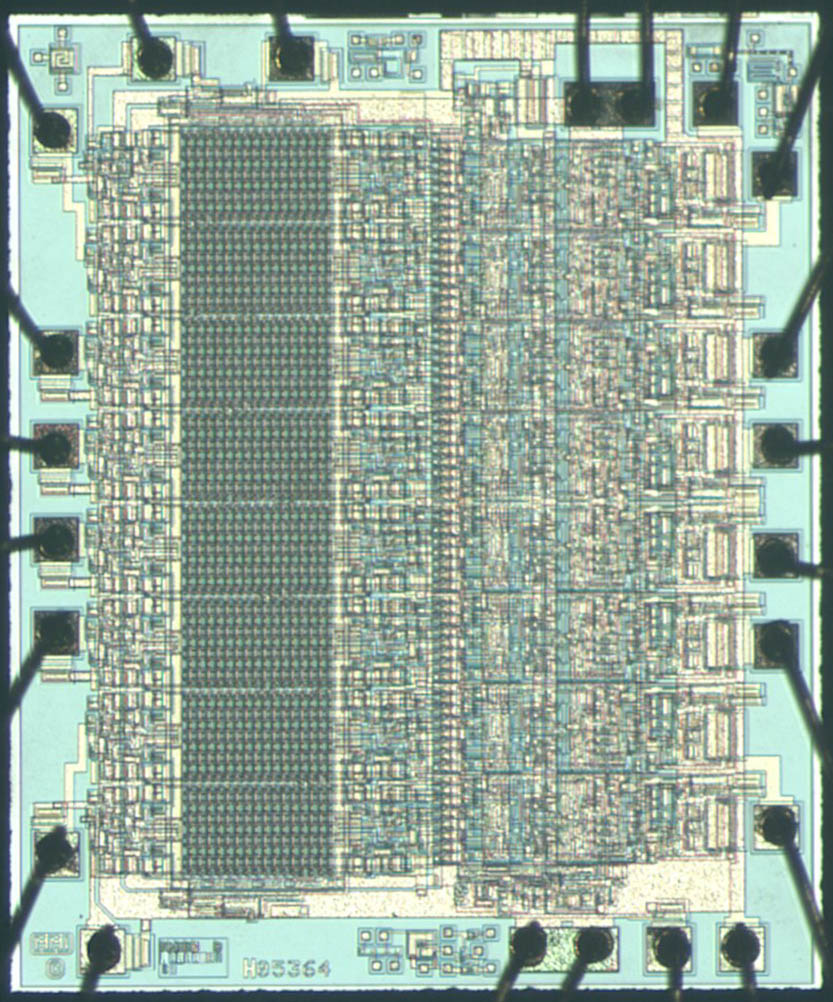 I was surprized how bad this next picture was. I was told by MEFAS that the image below was taken with a "stereo microscope". He said to get the image he got, use an "optical microscope". There is a metallurgy microscope at UAA that I will use to finish the project. 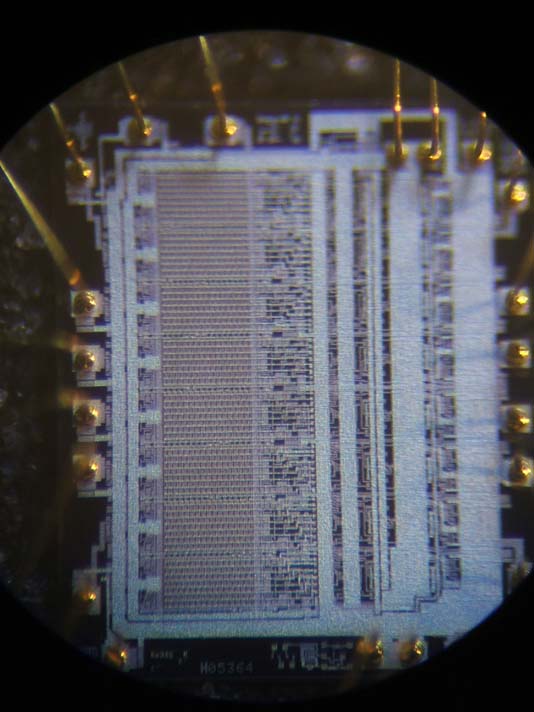 That's what I was able to get with the work microscope and a digital camera held up to the eye piece. Each block of the PAL should resemble this: 
Last edited by Grant Stockly; 09-12-2005 at 07:41 PM. 
|
|
|
 Similar Threads
|
||||
| Thread | Thread Starter | Forum | Replies | Last Post |
| QX5 microscope | tonigau | Electronics | 0 | 02-05-2006 05:09 PM |
| Microscope examination of a PLD | Grant Stockly | Electronics | 7 | 09-05-2005 02:56 PM |


 Threaded Mode